Mitochondria Structure: 4 Key Components From Double Membranes to Dynamic Cristae
The mitochondrion is more than the “powerhouse of the cell” — it’s an architectural marvel evolved from ancient symbiosis that bridges MITOCHONDRIA structure with function seamlessly. To understand its powerhouse role, you must first understand its sophisticated design.
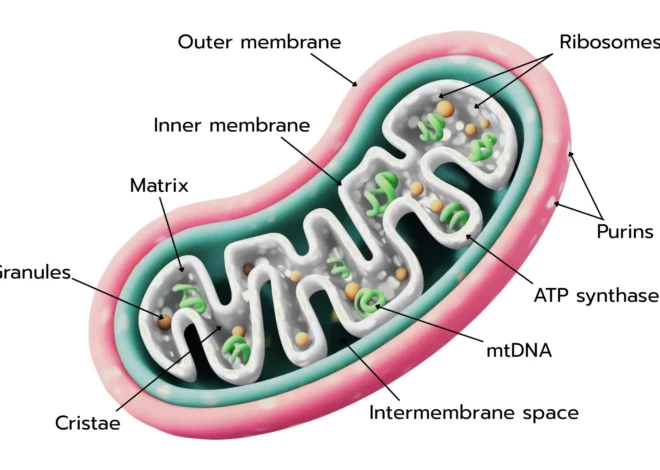
Core Structural Components
Each mitochondrion contains four major structural regions, each engineered for a specialized task.
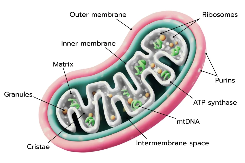
1. Outer Membrane
- Key Feature: Contains porins — also known as Voltage-Dependent Anion Channels (VDACs).
- Role: Acts like a selective sieve, allowing small molecules (under 5 kDa) to pass freely, while blocking larger proteins unless actively transported.
- Significance: Facilitates the free flow of ions, ATP, ADP, and metabolic intermediates.
- Source: NCBI – Mitochondrial Membranes
2. Inner Membrane
- Key Feature: Folded into intricate structures called cristae.
- Role: Houses the Electron Transport Chain (ETC) complexes I-IV and ATP synthase.
- Significance: These folds dramatically expand the surface area — up to 5–10 times more than a smooth membrane — allowing more space for ATP production machinery.
3. Intermembrane Space
- Key Feature: Narrow gap between inner and outer membranes.
- Role: Stores protons (H⁺) during electron transport, creating an electrochemical gradient.
- Significance: Contains cytochrome c, a vital protein for triggering apoptosis when the cell is damaged.
4. Matrix
- Key Feature: The innermost compartment, a gel-like space rich in enzymes and DNA.
- Role: Contains:
- mtDNA (Mitochondrial DNA)
- Mitochondrial ribosomes
- Enzymes for the Krebs cycle
- Calcium granules
- Significance: The matrix is the command center for mitochondrial genetics and fuel metabolism.
| Component | Main Molecules | Key Function |
| Outer Membrane | VDAC, porins | Molecule transport |
| Inner Membrane | ETC complexes, ATP synthase, cristae | ATP production |
| Intermembrane Space | Cytochrome c, H⁺ ions | Proton gradient, apoptosis |
| Matrix | mtDNA, ribosomes, Krebs cycle enzymes | Metabolism, genetic control |
Size and Shape
Mitochondria are typically 0.5–10 micrometers long. They aren’t static — they fuse, divide, and reshape themselves constantly to match the energy needs of the cell. This dynamic reshaping, via fission and fusion, maintains healthy mitochondria and removes damaged parts.
Source: Nature Reviews — Mitochondrial Dynamics
Linking Structure with Function
The real magic of mitochondria lies in how structural details drive function.
Cristae and Energy Efficiency
The cristae are not random folds — they are precision-engineered ridges packed with protein complexes.
- Heart cells: Use up to 40% of their cell volume for mitochondria to meet relentless energy demands.
- Cristae density: The denser the folds, the more ETC proteins can embed, leading to maximum ATP output.
- Supercomplexes: Complexes I-IV and ATP synthase often form supercomplex clusters on cristae to optimize electron flow and minimize energy loss.
| Cell Type | Cristae Density | ATP Demand |
| Cardiac Muscle | Extremely high | Constant contraction |
| Liver | High | Detoxification, metabolism |
| Neurons | High | Neurotransmission |
Source: ScienceDirect — Cristae Morphology
Contact Sites: Talking with Other Organelles
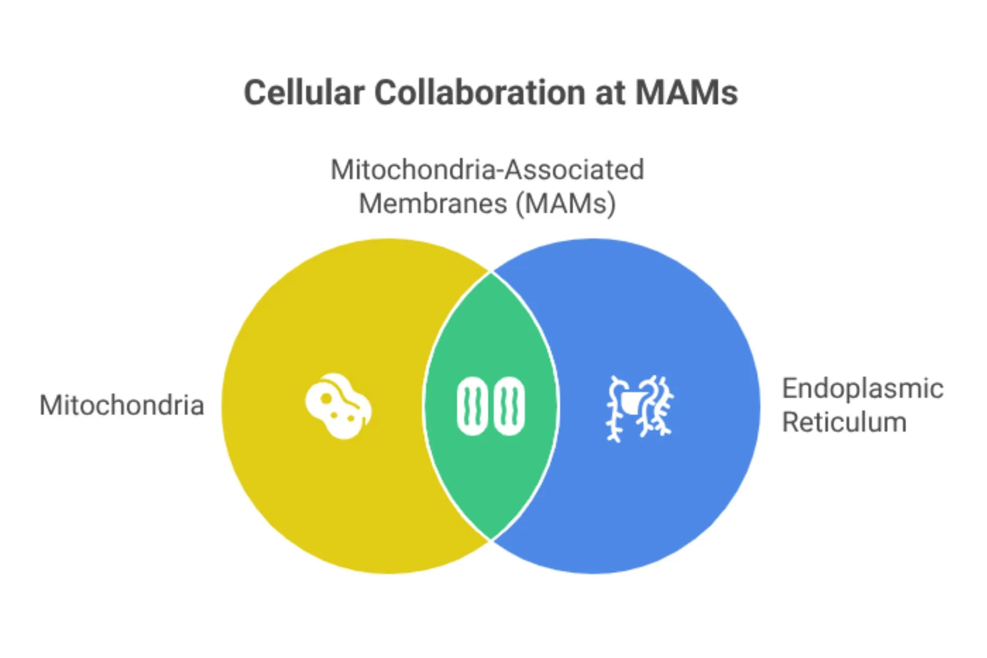
Mitochondria don’t work alone. They form contact sites called Mitochondria-Associated Membranes (MAMs) with the endoplasmic reticulum (ER). These sites:
- Regulate calcium exchange: Fine-tunes cellular signals for muscle contraction and hormone release.
- Facilitate lipid transfer: Crucial for membrane synthesis.
- Trigger apoptosis: If mitochondria detect damage beyond repair, they send death signals through MAMs.
Also read: Mitochondria Function Decoded
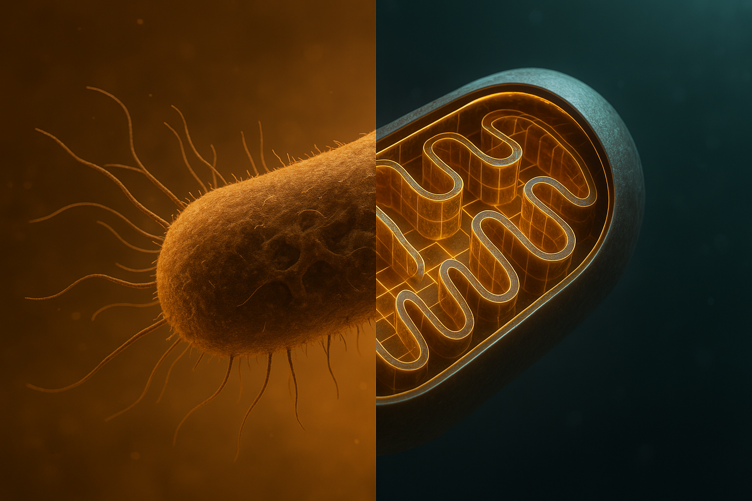
Evolutionary Roots and Unique mtDNA
Mitochondria evolved through endosymbiosis — an ancient free-living bacterium entered a larger cell and stuck around.
- mtDNA is bacterial-like: Circular, double-stranded, coding for 37 genes, mostly ETC proteins.
- Own Ribosomes: mtDNA has dedicated ribosomes for local protein synthesis, boosting efficiency.
- Location: Safely tucked inside the matrix, close to ETC machinery for direct use.
This bacterial ancestry explains why mitochondria divide like bacteria — independent of the cell cycle — and why their DNA is distinct from nuclear DNA.
Source: Cell — Endosymbiosis Theory
Diseases Linked to Structural Defects
When mitochondrial structures fail, the consequences are severe.
| Disease | Structural Defect | Impact |
| LHON (Leber’s Hereditary Optic Neuropathy) | Mutations in Complex I | Excessive ROS → optic nerve damage |
| Ischemic Heart Disease | Cristae damage → opens mPTP (Permeability Transition Pore) | Cytochrome c leaks → triggers cell death |
- ROS (Reactive Oxygen Species): Byproducts of ETC damage mtDNA and proteins.
- mPTP: A “fail switch” that releases apoptotic factors if stress is too high.
Source: PubMed — Mitochondrial Diseases
Modern Structural Research Techniques
Cryo-Electron Tomography
- Visualizes 3D structures of supercomplexes in near-native conditions.
- Reveals dynamic protein arrangements that standard microscopy misses.
Single-Mitochondrion Analysis
- Uses capillary electrophoresis to study individual mitochondria.
- Highlights heterogeneity — not all mitochondria in a cell are equal!
Source: Nature Methods — Cryo-ET
FAQs
Why does the inner membrane have folds (cristae)
Cristae folds multiply the inner membrane’s surface area by 5–10 times, allowing more ETC proteins and ATP synthase complexes to fit. This directly boosts a cell’s capacity to produce ATP, which is vital for high-energy tissues like the heart and brain. Without cristae, the energy output would plummet. Learn more
Where is mtDNA located and why does it mutate more?
mtDNA sits in the matrix, close to the ETC, which produces reactive oxygen species (ROS) as byproducts. These ROS can damage mtDNA, and since mitochondria lack robust repair systems like the nucleus, the mutation rate is 10–100 times higher. This is why aging and many degenerative diseases link back to mitochondrial mutations. Source
How do mitochondria change shape or move?
Mitochondria use fission (splitting) and fusion (joining) to maintain quality and energy distribution. Proteins like Drp1 pinch mitochondria apart during fission, while Mfn1/2 and OPA1 fuse them together. This dynamic reshaping lets mitochondria adapt to cellular stress and energy needs. Learn more
How is mitochondria structure different in cancer cells?
Cancer cells often have fewer cristae, prioritizing glycolysis over oxidative phosphorylation (Warburg effect). They may overexpress VDAC in the outer membrane to increase calcium influx, fueling rapid growth and resistance to cell death. Understanding these differences helps target mitochondrial metabolism in cancer therapy. Source
Conclusion
The mitochondrion’s genius lies in its structure: membranes, folds, spaces, and ancient DNA work in perfect unison to power life. By linking structure to function — from cristae surface area to dynamic fission/fusion — scientists decode diseases and design cutting-edge treatments.
Modern research pushes this frontier further, revealing mitochondria not just as static “powerhouses” but as living, changing machines at the heart of cellular life.